
LIFE IN PRISON...AGAINST TRUMP SUPPORTERS,,,MILLIONS IN FINES.....DEPORT HIS FAMILY,,,,LEFT MUST PAY
Uncovering the Truth: The Controversial FBI Interview That Exposes Ray Epps’ Role in the January 6th Capitol Riot
Amidst the chaos and confusion surrounding the events of January 6th at the US Capitol, one name has surfaced time and time again – Ray Epps. A figure shrouded in mystery, Epps has been the subject of intense speculation and scrutiny, with many questioning his true intentions on that fateful day.
And now, never-before-released information from an FBI interview has shed new light on Epps and his alleged involvement. But as the details emerge, one thing becomes clear – the conservative perspective on this revelation is one that cannot be ignored.
From the moment Epps was captured on camera urging protesters to "go into the Capitol," the public has been divided on his motives. Was he a true supporter of former President Trump, simply caught up in the fervor of the moment?
Or was he a plant, sent to incite violence and discredit the peaceful protests? The latter seems to be gaining traction as the FBI interview transcript reveals shocking information about Epps' background and actions.
Circles
Posts


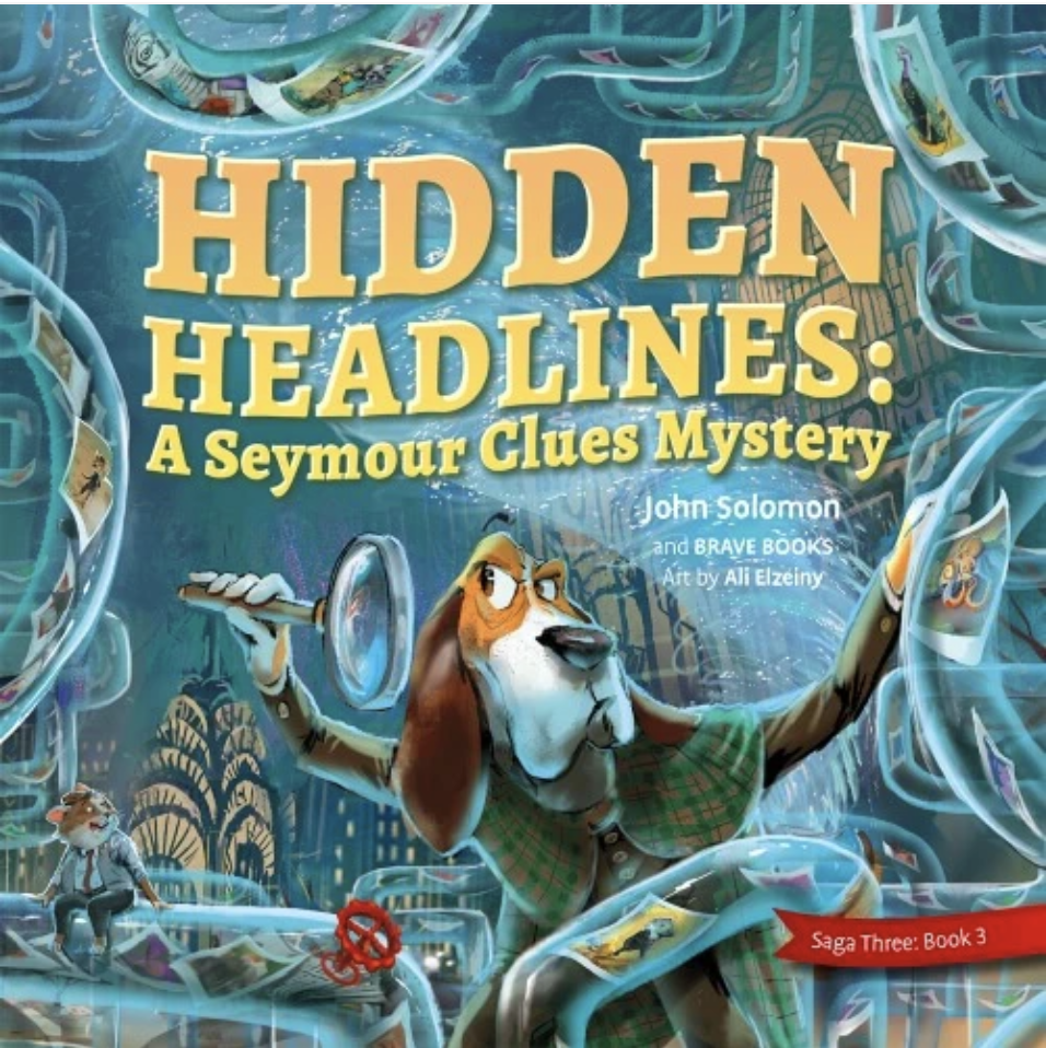
In John Solomon's book Hidden Headlines: A Seymour Clues Mystery, join Seymour Clues as he solves the mystery of who blocked the tubes around Starlotte City, keeping the Hamster Headlines newspaper from reaching its citizens. Along the way discover the importance of honesty, integrity, free speech, and the evils of intolerance and censorship.
Check it out here at Brave Books: https://bravebooks.us/?sca_ref=4492304.RjKJXiXAW3&utm_source=affiliate_program&utm_medium=uppromote&utm_campaign=cheryl-johnson
Support our patriotic products here: https://www.readwritetechnology.com/patriotic-products
FBI,,,CIA,,,DEMONICRAPS,,,MOB...DID IT,,,AND THERE STILL KENNEDIES. THAT RUN ON THE DEMONICRAP TICKET,,,WILL JOHN. FORGIVE THEM..
Revisiting the Shadows: A Conservative Analysis of JFK’s Assassination
The assassination of President John F. Kennedy remains one of the most pivotal moments in American history, shrouded in mystery and controversy. Despite the passage of time, the official narrative, as presented by the Warren Commission, has been met with skepticism and critical examination. Jerome Corsi and David Mantik, in their groundbreaking work, "The Assassination of John F. Kennedy: The Final Analysis," offer a compelling reevaluation of this tragic event, challenging the established account with new forensic evidence and analysis.
At the heart of their investigation is a meticulous examination of the autopsy X-rays of President Kennedy, which are preserved in the National Archives. Through their analysis, Corsi and Mantik present a startling conclusion: Kennedy was struck by three bullets to the head, with two entering from the front and one from the back at a lower angle. This finding is further supported by the independent review conducted by Dr. Michael Chesser, who examined the lateral X-ray film, corroborating the presence of multiple entry wounds.
Videos


Should parents be concerned about the hot-button mystery pneumonia, AKA “white lung syndrome,” affecting children in China? Is this a novel outbreak, or just another mechanism used by mainstream media to hide vaccine injuries?
Pediatrician Paul Thomas, M.D. weighs in about this phenomenon on #CHDTV.
“There’s no reason to worry. We’ve seen this before.”
“When you give a pharmaceutical or you vaccinate, there are often side effects. And then those side effects are never attributed to the vaccine or the pharmaceutical, they’re called a ‘syndrome.’”
Take Sudden Infant Death Syndrome (SIDS) for example. Rather than looking into the connection between SIDS and childhood innoculations, concerns raised by parents and doctors are instead mocked, silenced and ignored.
Maybe this is why no one is asking the million-dollar question: Were these kids recently administered a product from Big Pharma? Perhaps the answer is too much to bear.
Learn more about ‘White Lung Syndrome’ from two expert pediatricians on #CHDTV ?
https://live.childrenshealthdefense.org/chd-tv/shows/good-morning-chd/white-lung-syndrome-what-you-need-to-know/


On this episode on 360 View International Correspondent Roxana Solano explains how DNA has been used to solve crimes and mysteries across the globe. But who owns it? Can you own your DNA? Every time someone swabs for a 23andMe are they voluntarily forfeiting the right to their own molecules? Scottie Nell Hughes asks Criminal Defense Attorney, Joseph Tully, what should be done to protect the privacy of DNA?
Circles
Videos
Should parents be concerned about the hot-button mystery pneumonia, AKA “white lung syndrome,” affecting children in China? Is this a novel outbreak, or just another mechanism used by mainstream media to hide vaccine injuries?
Pediatrician Paul Thomas, M.D. weighs in about this phenomenon on #CHDTV.
“There’s no reason to worry. We’ve seen this before.”
“When you give a pharmaceutical or you vaccinate, there are often side effects. And then those side effects are never attributed to the vaccine or the pharmaceutical, they’re called a ‘syndrome.’”
Take Sudden Infant Death Syndrome (SIDS) for example. Rather than looking into the connection between SIDS and childhood innoculations, concerns raised by parents and doctors are instead mocked, silenced and ignored.
Maybe this is why no one is asking the million-dollar question: Were these kids recently administered a product from Big Pharma? Perhaps the answer is too much to bear.
Learn more about ‘White Lung Syndrome’ from two expert pediatricians on #CHDTV ?
https://live.childrenshealthdefense.org/chd-tv/shows/good-morning-chd/white-lung-syndrome-what-you-need-to-know/
On this episode on 360 View International Correspondent Roxana Solano explains how DNA has been used to solve crimes and mysteries across the globe. But who owns it? Can you own your DNA? Every time someone swabs for a 23andMe are they voluntarily forfeiting the right to their own molecules? Scottie Nell Hughes asks Criminal Defense Attorney, Joseph Tully, what should be done to protect the privacy of DNA?
Posts
LIFE IN PRISON...AGAINST TRUMP SUPPORTERS,,,MILLIONS IN FINES.....DEPORT HIS FAMILY,,,,LEFT MUST PAY
Uncovering the Truth: The Controversial FBI Interview That Exposes Ray Epps’ Role in the January 6th Capitol Riot
Amidst the chaos and confusion surrounding the events of January 6th at the US Capitol, one name has surfaced time and time again – Ray Epps. A figure shrouded in mystery, Epps has been the subject of intense speculation and scrutiny, with many questioning his true intentions on that fateful day.
And now, never-before-released information from an FBI interview has shed new light on Epps and his alleged involvement. But as the details emerge, one thing becomes clear – the conservative perspective on this revelation is one that cannot be ignored.
From the moment Epps was captured on camera urging protesters to "go into the Capitol," the public has been divided on his motives. Was he a true supporter of former President Trump, simply caught up in the fervor of the moment?
Or was he a plant, sent to incite violence and discredit the peaceful protests? The latter seems to be gaining traction as the FBI interview transcript reveals shocking information about Epps' background and actions.
In John Solomon's book Hidden Headlines: A Seymour Clues Mystery, join Seymour Clues as he solves the mystery of who blocked the tubes around Starlotte City, keeping the Hamster Headlines newspaper from reaching its citizens. Along the way discover the importance of honesty, integrity, free speech, and the evils of intolerance and censorship.
Check it out here at Brave Books: https://bravebooks.us/?sca_ref=4492304.RjKJXiXAW3&utm_source=affiliate_program&utm_medium=uppromote&utm_campaign=cheryl-johnson
Support our patriotic products here: https://www.readwritetechnology.com/patriotic-products
FBI,,,CIA,,,DEMONICRAPS,,,MOB...DID IT,,,AND THERE STILL KENNEDIES. THAT RUN ON THE DEMONICRAP TICKET,,,WILL JOHN. FORGIVE THEM..
Revisiting the Shadows: A Conservative Analysis of JFK’s Assassination
The assassination of President John F. Kennedy remains one of the most pivotal moments in American history, shrouded in mystery and controversy. Despite the passage of time, the official narrative, as presented by the Warren Commission, has been met with skepticism and critical examination. Jerome Corsi and David Mantik, in their groundbreaking work, "The Assassination of John F. Kennedy: The Final Analysis," offer a compelling reevaluation of this tragic event, challenging the established account with new forensic evidence and analysis.
At the heart of their investigation is a meticulous examination of the autopsy X-rays of President Kennedy, which are preserved in the National Archives. Through their analysis, Corsi and Mantik present a startling conclusion: Kennedy was struck by three bullets to the head, with two entering from the front and one from the back at a lower angle. This finding is further supported by the independent review conducted by Dr. Michael Chesser, who examined the lateral X-ray film, corroborating the presence of multiple entry wounds.
https://www.chat-hozn3.com/blogs/5022/a-simple-key-for-slot-gacor-777-unveiled
https://www.chat-hozn3.com/blogs/5023/winsgoal-no-further-a-mystery
https://www.chat-hozn3.com/blogs/5024/daftar-slot-gacor-777-fundamentals-explained
https://www.chat-hozn3.com/blogs/5025/5-easy-facts-about-situs-slot-gacor-777-described
https://www.chat-hozn3.com/blogs/5026/not-known-facts-about-situs-slot-gacor-777
https://www.chat-hozn3.com/blogs/5027/getting-my-winsgoal-to-work-slot-gacor-777-online
https://www.chat-hozn3.com/blogs/5028/slot-gacor-777-for-dummies
https://www.chat-hozn3.com/blogs/5029/how-slot-gacor-777-can-save-you-time-stress-and
https://www.chat-hozn3.com/blogs/5030/not-known-facts-about-daftar-slot-gacor-777
https://www.chat-hozn3.com/blogs/5031/rumored-buzz-on-daftar-slot-gacor-777
https://www.chat-hozn3.com/blogs/5032/indicators-on-winsgoal-you-should-know
https://www.chat-hozn3.com/blogs/5033/slot-gacor-777-hari-ini-no-further-a-mystery
https://www.chat-hozn3.com/blogs/5034/5-essential-elements-for-situs-slot-gacor-777
https://www.chat-hozn3.com/blogs/5035/top-latest-five-slot-gacor-777-urban-news
https://www.chat-hozn3.com/blogs/5036/a-review-of-winsgoal-daftar-slot-gacor-777
https://www.chat-hozn3.com/blogs/5037/slot-gacor-777-hari-ini-no-further-a-mystery
https://www.chat-hozn3.com/blogs/5038/the-smart-trick-of-link-slot-gacor-777-that-no
https://www.chat-hozn3.com/blogs/5040/a-review-of-link-slot-gacor-777
https://www.chat-hozn3.com/blogs/5039/a-review-of-link-slot-gacor-777
https://www.chat-hozn3.com/blogs/5041/how-much-you-need-to-expect-you-ll-pay-for
https://www.chat-hozn3.com/blogs/5042/slot-gacor-777-hari-ini-no-further-a-mystery
https://www.chat-hozn3.com/blogs/5043/a-review-of-winsgoal-daftar-situs-slot-gacor-777
https://www.chat-hozn3.com/blogs/5044/slot-gacor-777-hari-ini-no-further-a-mystery
https://www.chat-hozn3.com/blogs/5045/examine-this-report-on-situs-slot-gacor-777-hari-ini
https://www.chat-hozn3.com/blogs/5046/slot-gacor-777-fundamentals-explained
https://www.chat-hozn3.com/blogs/5047/not-known-facts-about-situs-slot-gacor-777
https://www.chat-hozn3.com/blogs/5048/winsgoal-no-further-a-mystery-of-situs-slot-gacor-777
https://www.chat-hozn3.com/blogs/5049/not-known-facts-about-situs-slot-gacor-777
https://www.chat-hozn3.com/blogs/5050/5-easy-facts-about-link-slot-gacor-777-described
https://www.chat-hozn3.com/blogs/5051/rumored-buzz-on-slot-gacor-777-hari-ini
https://www.chat-hozn3.com/blogs/5052/the-5-second-trick-for-winsgoal-situs-slot-gacor-777
https://www.chat-hozn3.com/blogs/5053/examine-this-report-on-slot-gacor-777-hari-ini
https://www.chat-hozn3.com/blogs/5054/top-latest-five-
https://www.chat-hozn3.com/blogs/5055/5-simple-techniques-for-winsgoal-situs-slot-gacor-777
https://www.chat-hozn3.com/blogs/5056/slot-gacor-777-hari-ini-no-further-a-mystery
https://www.chat-hozn3.com/blogs/5057/new-step-by-step-map-for-situs-slot-gacor-777
https://www.chat-hozn3.com/blogs/5058/top-latest-five-daftar-slot-gacor-777-urban-news
https://www.chat-hozn3.com/blogs/5059/everything-about-slot-gacor-777
https://www.chat-hozn3.com/blogs/5060/5-essential-elements-for-situs-slot-gacor-777
https://www.chat-hozn3.com/blogs/5061/a-review-of-link-slot-gacor-777

ANATOMY OF A FALL , a great sociology study film in what it feels like to suddenly find yourself being accused of committing a murder just because it looks that way to the authorities. This is a film made by a director who loves film making. You get completely dragged into the life of the characters. This is a thinking person's movie.
10/10 "go sees" #anatomyofafall #movie #film #review #drama #guess #mystery #mysterymovie https://www.youtube.com/watch?v=FUXawkH-ONM

For the past year, Sandra, her husband Samuel, and their eleven-year-old son Daniel have lived a secluded life in a remote town in the French Alps. When Samu...
![]() www.youtube.com
www.youtube.com